
Online POCUS Training,
Tailored to Your Specialty
Transform your bedside practice with online point-of-care
ultrasound (POCUS) training customized to your specialty.
ultrasound (POCUS) training customized to your specialty.

Who We Serve
Individuals
Physicians, fellows, APPs, residents, and medical students—enhance your POCUS skills with training geared toward your specialty. Learn at your own pace, earn CME credits or certificates of completion, and gain confidence in scanning at the bedside.
Groups and Residency Programs
Residency directors and educators—give your learners the structured, comprehensive training they need with our custom-built residency curriculum. Save on faculty time, offer specialty-specific modules, and shape a new generation of POCUS leaders.
What Makes Us Different
Case of the Month
Write your awesome label here.
Tip of the Day
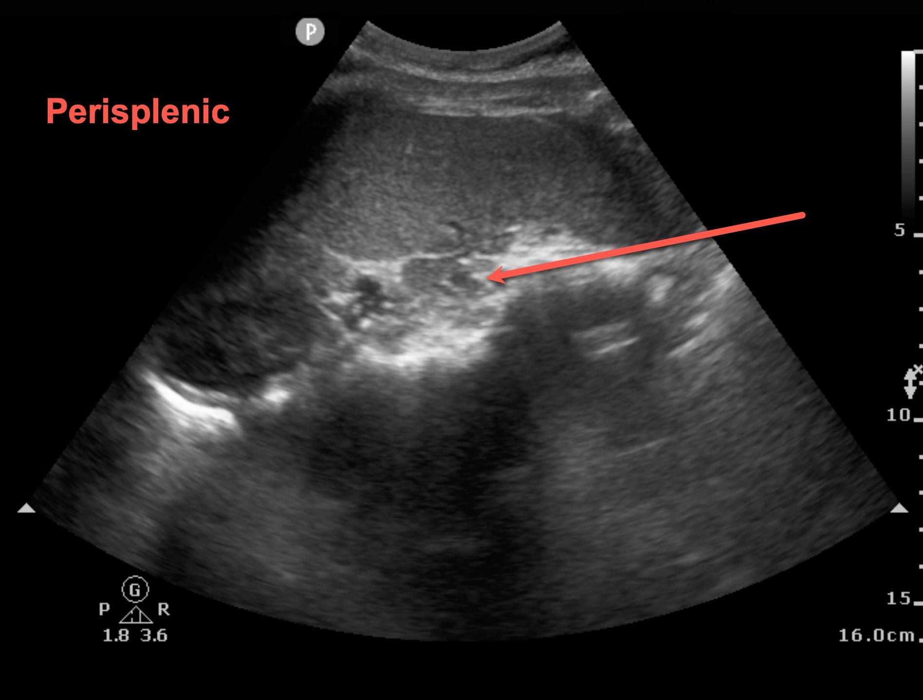
Featured Image
Case of the Month
Write your awesome label here.
Journal Article
Tip of the Day
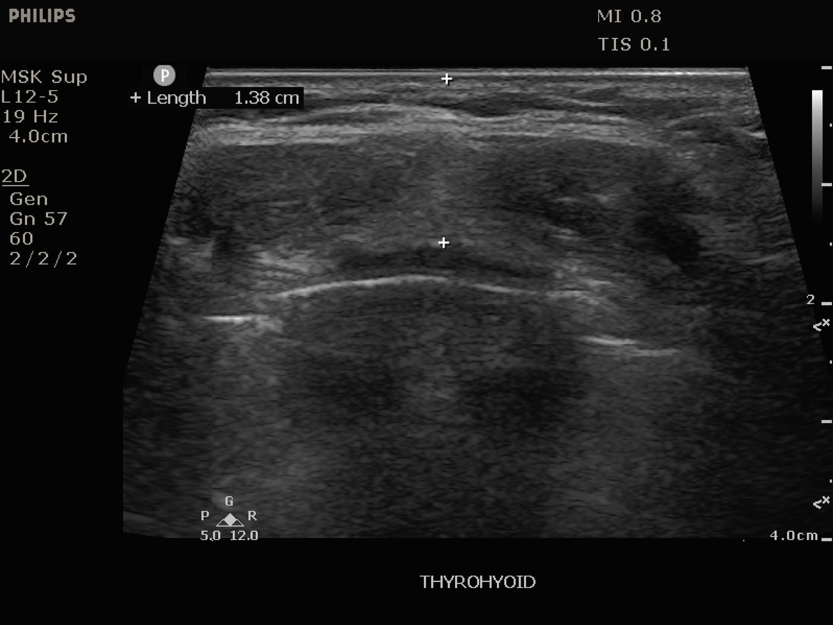
Featured Image
Case of the Month
Write your awesome label here.
Tip of the Day
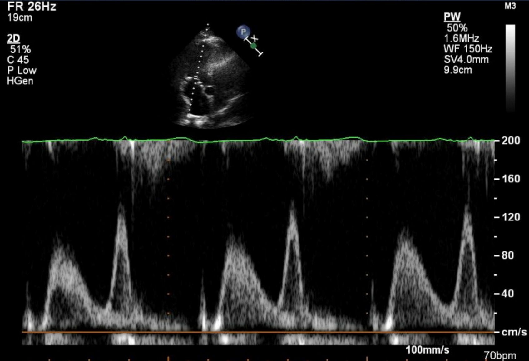
Featured Image
Case of the Month
Write your awesome label here.
Journal Article
Tip of the Day
Featured Image
Case of the Month
Write your awesome label here.
Tip of the Day
Featured Image
Case of the Month
Write your awesome label here.
Tip of the Day
Featured Image
Case of the Month
Write your awesome label here.
Journal Article
Tip of the Day
Featured Image
Featured Image
Featured Image
Write your awesome label here.
Elevate Your POCUS Skills
Choose your specialty and start learning—one focused POCUS course at a time.

Copyright © 2025